Fig. 1.
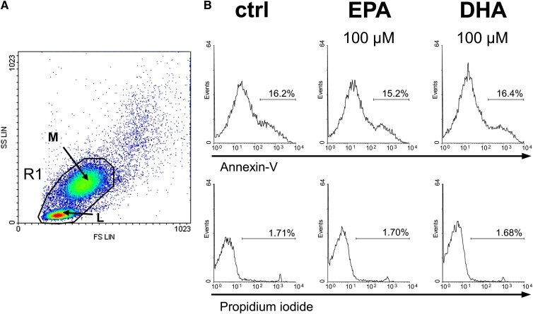
EPA and DHA do not affect cell viability. Cell viability was flow-cytometrically assessed by annexin-V and propidium iodide exclusion double staining. (A) Forward scatter (FS) against side scatter (SS) dot plot of human PBMC after alloreactive stimulation. Data acquisition was set to lymphocytes (L) and monocytes (M) within the scatter gate R1 of 50,000 total counts. (B) Representative histograms of fluorescence intensities of PBMC treated without or with 100 µM fatty acid for 24 h. Relative to the control (ctrl), annexin-V-positive and PI-negative cells were defined as early apoptotic cells; annexin-V-positive and PI-positive cells were defined as late apoptotic and necrotic cells.

