
Fig. 4.
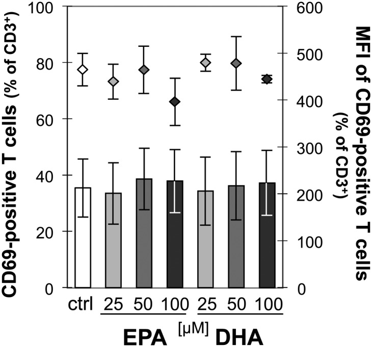
EPA and DHA do not affect CD69 expression on activated T cells. PBMC were incubated for 19 h without or with increasing concentrations of EPA or DHA before stimulation with ConA for subsequent 5 h. Cells were then stained for CD3 and CD69. Expression of cell surface markers was flow-cytometrically analyzed. Data are expressed as means ± SD of n = 3. Right scales denote mean fluorescence intensity (MFI) depicted as dots.