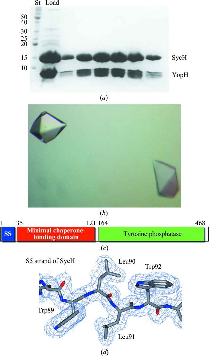
Figure 1.
Purification, crystallization and electron density of YopH–SycH. (a) Coomassie-stained SDS–PAGE analysis of the purified YopH–SycH complex used for crystallization. Lane St contains protein standard markers for assigning molecular weight (labeled in kDa). (b) Well diffracting crystals of the YopH21–63–SycH complex. (c) Domain schematic of YopH. (d) 2F o − F c model-phased electron-density maps contoured at 1σ shown in blue with the final refined model shown.

