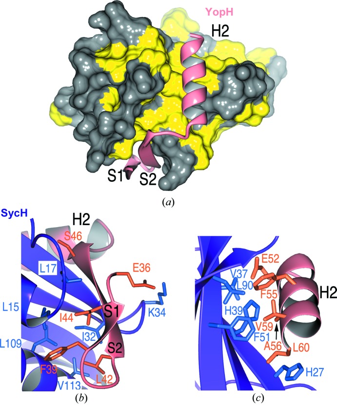
Figure 4.
YopH–SycH interactions. (a) Hydrophobic patches in SycH (yellow) that bind the YopH polypeptide (shown as a ribbon cartoon in salmon). (b) Residues making contact between the S2 strand (β-motif) of YopH (salmon and orange) and SycH (purple and blue). (c) Contacts between the YopH H2 helix and the SycH helix-binding groove.

