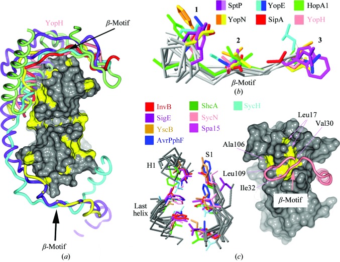
Figure 5.
YopH possesses a canonical β-motif. (a) Comparison of T3SS effector nonglobular polypeptides from several complexes with chaperones (the alignment was generated by aligning the complexes based on the conserved folds of the chaperones). Effector polypeptides include Salmonella SipA (PDB entry 2fm8, red; Lilic et al., 2006 ▶) and SptP (PDB entry 1jyo, purple; Stebbins & Galán, 2001b ▶), Yersinia YopN (PDB entry 1xkp, ellow; Schubot et al., 2005 ▶) and YopE (PDB entry 1l2w, cyan; Birtalan et al., 2002 ▶) and Pseudomonas syringae HopA1 (PDB entry 4g6t, green; C. E. Stebbins, R. Janjusevic & C. M. Quezada, unpublished work). Placed roughly for orienting the image is a surface rendering of SycH with hydrophobic patches shown in yellow. (b) Alignment of several β-motifs from animal and plant effectors. (c) Alignment of the β-motif binding pocket in chaperones from animal and plant pathogens. On the left are three key elements of structure in all chaperones characterized to date, consisting of six highly conserved hydrophobic amino acids which closely superimpose and which frequently make contacts with β-motif residues. On the right is a space-filling representation of SycH with the six conserved hydrophobic residues of the chaperone pocket shown in yellow and indicated by arrows. The main-chain trace of YopH is drawn as a tube in salmon; the β-motif is indicated.

