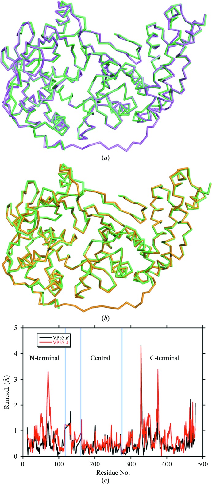
Figure 3.
Backbone structural comparison of the VP55 monomer with VP55 from the heterodimer. (a) Overlay of VP55 chain A (magenta) on PDB entry 2ga9 (green). (b) Overlay of VP55 chain B (yellow) on PDB entry 2ga9 (green). The N-terminal domain of VP55 is shown on the right-hand side, the C-terminal domain is shown on the left and the loop (118–129) is at the bottom. (c) R.m.s. deviations: VP55 chain A versus PDB entry 2ga9, red; VP55 chain B versus PDB entry 2ga9, black.

