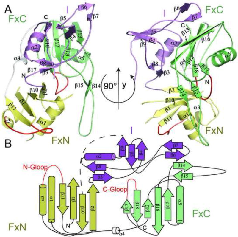
Figure 1.
Structure and topology of Cmr3 when bound to Cmr2dHD. (A) Secondary and domain structures of Cmr3 in two orientations. “FxN” and “FxC” refer to the Ferredoxin folds of the N-terminal and C-terminal domains and “I” refers to the insertion domain. Cmr3 domains are shown in different colors (FxN: Yellow, FxC: Light green, I: purple) (B) Topology of the Cmr3 structure. The two glycine-rich loops are shown in red. “N-Gloop” refers to the glycine-rich loop of the N-terminal domain and “C-Gloop” refers to the glycine-rich loop of the C-terminal domain.

