
Figure 2.
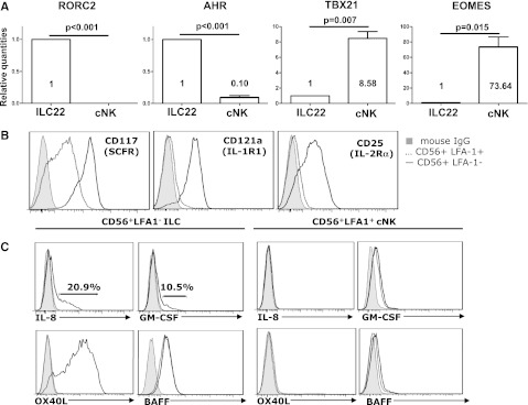
Transcription factors, cytokine receptors, and other molecules expressed in human IL-22–producing ILCs. (A) Expression of RORC2, AHR, TBX21, and Eomes was measured by real-time qPCR from HSC-derived cNK cells (CD56+CD94+CD7+/−CD117lowLFA-1+) and ILC22 cells (CD56+CD94−CD7−CD117highLFA-1+) after sorting. In ILC22 cells AHR and RORγt are highly expressed but are lower-expressed or absent in cNK cells. T-bet and Eomes expression is higher in CD56+LFA-1+ cNK cells. Transcripts in cNK cells were used as reference samples for relative quantification in ILC22 (ΔΔCT method, n = 3). (B) HSC-derived CD56+LFA-1− ILC22 but not cNK cells show expression of IL-1R1 and CD25 and CD117. (C) In addition to IL-22, CD56+LFA-1− ILC22 cells (left) express IL-8, GM-CSF (intracellular), and OX40 ligand (surface) when stimulated with IL-1β+23 (10 ng/ml each for 6 hours). Intracellular BAFF expression is detected in CD56+LFA-1− ILC22 cells without cytokine stimulation. All of the above were not detected on cNK cells (right). Gray-filled histograms are mouse immunoglobulin G, dotted lines are unstimulated, and solid lines are stimulated. Results are representative of 10 donors. IgG, immunoglobulin G.