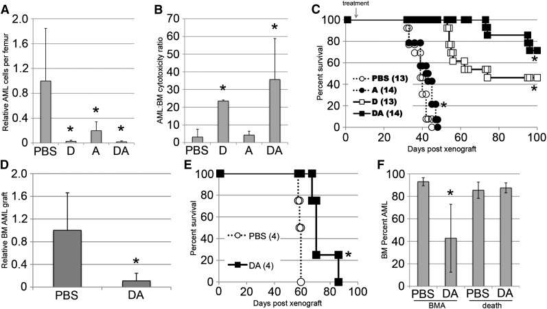
Figure 3.
Chemotherapy shows efficacy against human AML in vivo. (A) Human MA9-NRas AML cells remaining in the femurs of mice treated with chemotherapy were quantified on day 8 by cell counts and flow cytometry. The average AML cell number for the PBS group (1000 to 4000 cells per femur) was set to 1.0 in each experiment. (B) By using these data and data from Figure 2H, a ratio of AML to normal BM toxicity was calculated. (C) Mice engrafted and treated as in (A) were followed for survival. (D) Mice were injected with an AML patient sample (AMLCC2) and treated with chemotherapy. BM grafts were determined 6 to 8 weeks later. The average PBS grafts (15% to 40% AML) were set to 1.0 for each experiment for normalization. (E) Mice engrafted with a second AML patient sample (AMLCC1) were followed for survival. (F) BM grafts of AMLCC1-engrafted mice were determined at day 45 and again at time of death for each mouse. *(A, B, D, F) indicates P < .05 by the Student t test. *(C, E) indicates P < .05 by the log-rank test.

