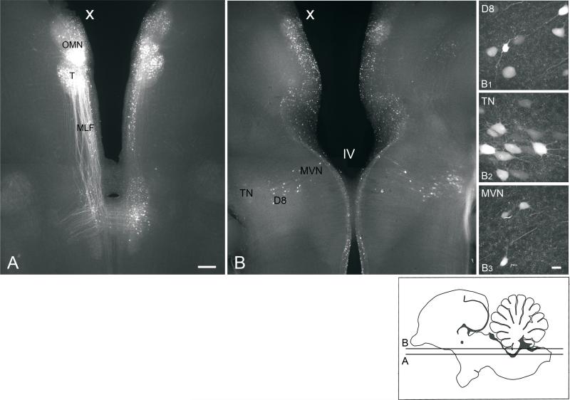
Fig. 3.
A, B, Low power, conventional fluorescence images (4x objective) of 300 μm, horizontal sections prepared from an E13 brainstem preparation, which received a unilateral extracellular biocytin injection into the oculomotor nucleus (X, injected side). Section B is dorsal to section A. B1-B3, high-power confocal images (40x objective) of retrogradely labeled neurons in the ipsilateral descending vestibular nucleus (D8), contralateral tangential nucleus (TN), and ipsilateral MVN, respectively, which appear in low power in B. The focal plane for B1-B3 may differ from each other and B. Insert, diagram of sagittal section of the brainstem indicating the levels of A and B. Scale bar in A, 200 μm, also refers to B. Scale bar in B3, 25 μm, also refers to B1 and B2. All photomicrographs are oriented with anterior to the top.

