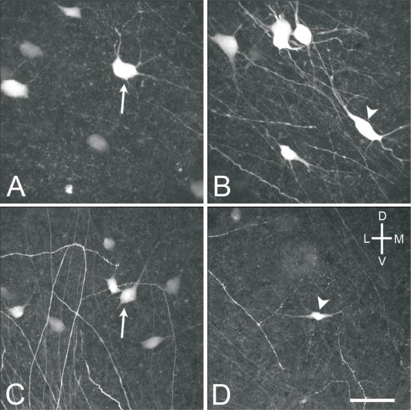
Fig. 7.
High power (40x objective) confocal images of MVNVL stellate (arrows) and elongate (arrowheads) neurons from E16 brainstem preparations. A, Stellate cell labeled after injecting the contralateral oculomotor nucleus. B, Elongate cell labeled after injecting the contralateral oculomotor nucleus. C, Stellate cell labeled after injection the contralateral abducens nucleus. D, Elongate cell labeled after injecting the ipsilateral abducens nucleus. Scale bar, 50 μm.

