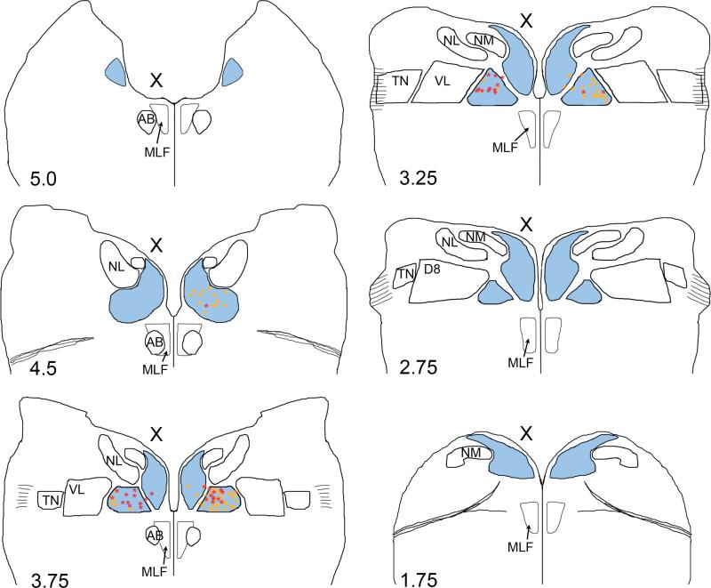
Fig. 9.
Summary diagram of brainstem sections showing the locations of retrogradely labeled MVN neurons after brainstem injections into the oculomotor (yellow squares, n=3 animals) or abducens nucleus (red stars, n=3 animals) at E13. The anteroposterior levels of the brainstem sections are labeled with reference to Fig. 2. Outlines are arranged from anterior (top left) to posterior (bottom right). To correct for tilt in the section plane, the level of each side of the section was evaluated independently to determine the anteroposterior level. The injected side is indicated (X). MVN (blue); NM, nucleus magnocellularis; NL, nucleus laminaris, TN, tangential nucleus; D8, descending vestibular nucleus; VL, ventrolateral vestibular nucleus, MLF, medial longitudinal fasciculus.

