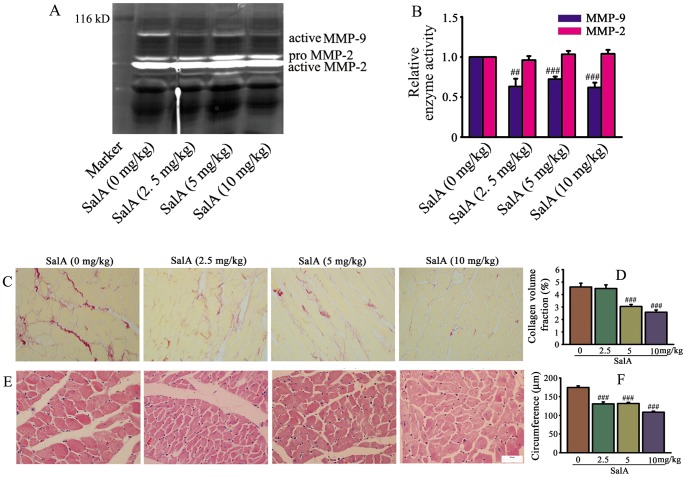
Figure 7. SalA inhibited MMP-9 activity and ameliorated cardiac remodeling in SHRs.
(A) The representative zymogram for MMP-9 and MMP-2 enzymatic activities in each treatment group in vivo. (B) Quantification of MMP-9, MMP-2 activity expressed as fold decrease versus SalA (0 mg/kg). (C) Representative area of whole heart stained by Sirius red. The position of collagen deposition was stained in red. (D) Quantitative data of collagen volume fraction for C. (E) Representative images of heart stained by haematoxylin and eosin. (F) Quantification of cardiomyocyte circumference with different treatment for E. Results are expressed as mean±S.E. #<0.05, ##p<0.01, ###p<0.001 versus SalA (0 mg/kg).

