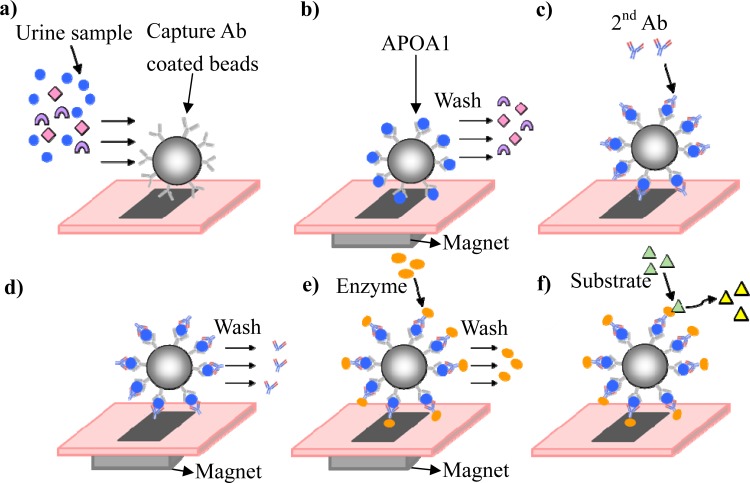
Figure 2.
Illustration of the working principles behind the bead-based ELISA using the microfluidic chip. (a) A urine sample containing the target protein and antibody-coated magnetic beads was introduced into the chip. (b) After the incubation process, the target protein was captured, and the unwanted protein was washed out. (c) and (d) The secondary antibody was bound to the antigen, and the excess antibody was washed out. (e) and (f) The enzyme was linked to the secondary antibody, and after the excess enzyme was washed out, a substrate was used to quantitatively measure the target protein.

