
Figure 4. Confocal laser scanning microscopy analysis of splenocytes co-cultured with PS-conjugated antigens.
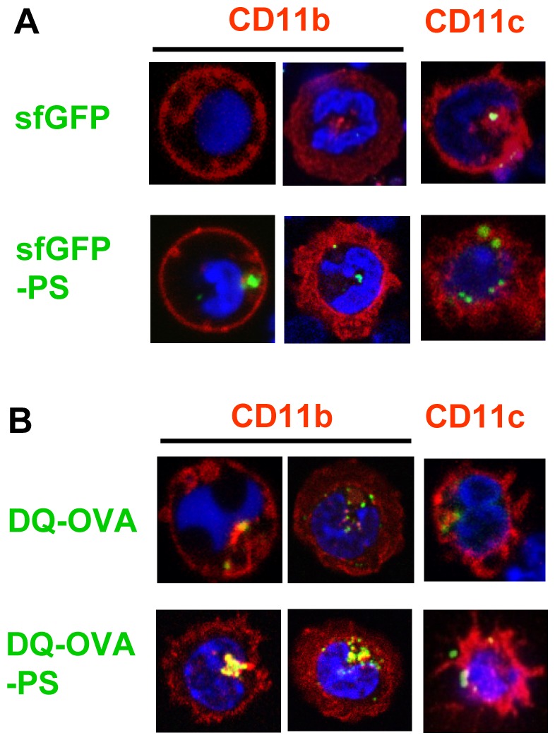
(A, B) CD11b+ or CD11c+ cells were cultured with sfGFP, sfGFP-PS, DQ-OVA or DQ-OVA-PS plus Hoechst33342 for 60 min at 37°C. After the incubation, cells were washed with PBS, and then analyzed under a LSM780 confocal laser scanning microscope system. Blue: cell nucleus, Green: sfGFP or DQ-OVA, Red: CD11b or CD11c.