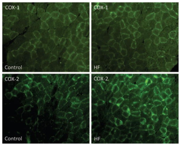
Figure 1. Immunostaining of COX-1 and COX-2.
Immunofluorescence methods were used to examine expression of cyclo-oxygenase (COX) isoforms COX-1 and COX-2 within the hindlimb muscles of control rats and rats with heart failure (HF rats). Top panels are typical images showing that COX-1 appears in the hindlimb muscles; the similar fluorescence staining is observed in control and HF rats. Bottom panels are typical immunofluorescence images showing that COX-2 is localized in the hindlimb muscles and that greater fluorescence staining is observed in HF than in control rats.

