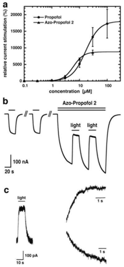
Figure 3.

a) α1β2γ2 GABAA receptors were expressed in Xenopus oocytes. Currents were activated with a concentration of GABA eliciting 0.3 % of the maximal current amplitude (EC0.3) without or with increasing amounts of either propofol or AP2. Mean ± SEM of four experiments is shown. b) 1 μM GABA was applied repetitively until a stable current response was observed. Co-application of 1.5 μM AP2 with GABA which resulted in current potentiation. During co-application, the oocyte was exposed to a light source emanating 360–370 nm light. As a consequence, current stimulation rapidly decreased until it reached a steady level. When the light-source was turned off, the amplitude increased again. This procedure was repeated. This experiment was repeated independently six times using different oocytes. c) α1β2γ2 GABAA receptors were expressed in HEK-cells. 0.5 μM GABA was co-applied with 5 μM AP2. Subsequently, the perfusion was stopped and the cells were exposed to the light source. The inward current amplitude decreased rapidly and increased again upon turning off the light-source (trace left). Current decrease (trace top right) and increase (trace bottom right) were each fitted with a mono-exponential function with τ amounting to 1.1 ± 0.4 s (n = 7) and 2.0 ± 0.7 s (n = 6), respectively.
