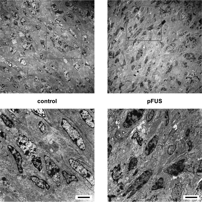
Figure 3.
(Color online) Representative transmission electron micrographs of control and pFUS treated tumors. Bottom images are expanded from boxes in top images. Tumors exposed to pFUS showed pervasive widening of intercellular spaces (arrows) between cells in the parenchyma, which were absent in the untreated tumors (scale bar = 5 μm).

