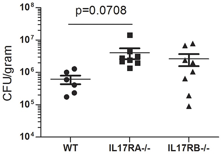
Figure 2. Colonization of IL-17RB deficient mice with H. pylori.

IL-17RA−/− mice, IL-17RB−/− mice and WT mice were infected with H. pylori strain SS1. Levels of colonization were measured by plating serial dilutions of stomach homogenates. The number of colony forming units (CFU) was then calibrated to the weight of the tissue and CFU/gram is presented on the graphs for 3 months post-infection.
