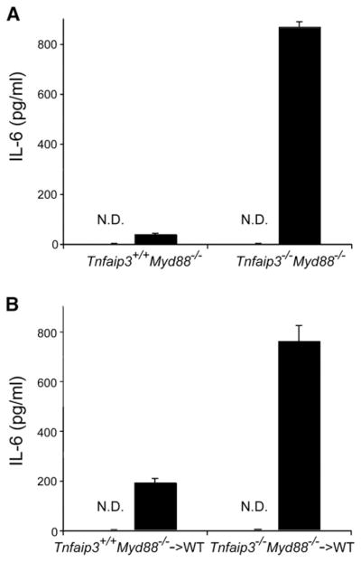
Figure 6. A20 Restricts MDP-Induced Inflammatory Responses In Vivo.

(A and B) ELISA analyses of serum from MDP-injected mice. Twenty-five milligrams over kilograms of MDP (or H2O) was injected into (A) intact Tnfaip3+/+ Myd88−/− and Tnfaip3−/− Myd88−/− mice, and (B) chimeric mice reconstituted with HSCs from Tnfaip3+/+ Myd88−/− and Tnfaip3−/− Myd88−/− mice. Serum was harvested 4 hr after MDP injection and analyzed for IL-6 levels by ELISA. White columns indicate samples from mice injected with H2O, and indicate that no IL-6 above baseline was detected (ND). Black columns indicate samples from MDP-injected mice. Note increased levels of serum IL-6 in (A) intact Tnfaip3−/− Myd88−/− mice compared with Tnfaip3+/+ Myd88−/− mice and in (B) chimeric mice bearing Tnfaip3−/− Myd88−/− HSCs compared with those bearing Tnfaip3+/+ Myd88−/− cells. Data were obtained from three sets of paired mice.
