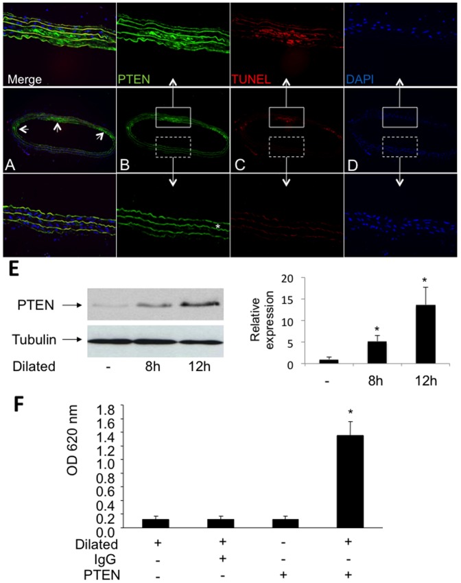
Figure 1. PTEN expression and apoptotic cells in vivo.
A–D, Immunoreactivity of PTEN (green), apoptotic smooth muscle cells (TUNEL staining, red) and nuclei (DAPI, blue) is shown in representative sections of an injured rat carotid artery 12 h after balloon injury. The autoflourescence of the elastic laminae (* in figure 1B) allows the identification of the media. Strongly injured areas of the dilated vessel were identified by a reduced cellularity and by the accumulation of apoptotic cells. Higher magnifications of vessel areas which fulfill these criteria are shown in the upper panel whereas higher magnifications of less injured vessel areas are shown in the lower panel. E PTEN expression is upregulated in dilated rat carotid arteries in vivo as determined by western blotting using a specific PTEN antibody. Detection of β-tubulin served as loading control. The relative expression of PTEN as determined by densitometric analysis of immunoblots is shown (n = 3; *P<0.05). F, The activity of PTEN is enhanced after vascular injury. Shown is the phosphatase activity as determined by immunoprecipitated PTEN from lysates of dilated and undilated rat carotid arteries. Immunoprecipitations employing an IgG iso-antibody and without addition of any antibodies served as controls. Results are expressed as mean OD650 ± SD (*P<0.001, n = 4).

