
Figure 2. PTEN expression in human coronary VSMC in vitro.
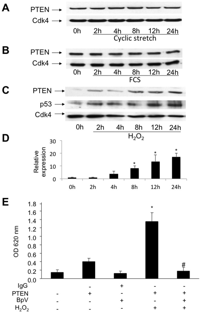
A, PTEN-expression is not upregulated by mechanical stress in VSMC. Lysates of cells exposed to mechanical forces using a stretching device were analyzed by western blotting using specific antibodies. B, Lysates of SMC exposed to growth medium (FCS). PTEN expression was not upregulated after 24 h. Protein expression was determined using specific antibodies. Detection of Cdk4 served as loading control. C, Lysates of SMC exposed to oxidative stress using 500 µM H2O2. PTEN upregulation was triggered by oxidative stress within 24 h. Detection of p53 and Cdk4 served as apoptotic marker and loading control, respectively. D, The upregulation of PTEN protein levels was quantified by densitometric analysis of immunoblots (n = 3; *P<0.05). E, The upregulation of PTEN activity is mediated by H2O2-induction. Shown is a phosphatase activity assay of immunoprecipitated protein from lysates of HcASMC with and without 24 h H2O2–treatment. Immunoprecipitations from lysates employing an IgG iso-antibody without H2O2-treatment with and without bpV supplementation, an anti-PTEN-antibody without H2O2 and bpV treatment and an anti-PTEN-antibody with H2O2– and bpV-treatment served as controls. Results are expressed as mean OD650 ± SD using an ELISA plate reader (# P<0.001, *P<0.001; n = 4).