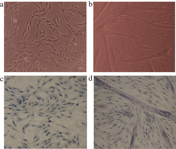
Figure 5.
Microscopic view of proliferating and differentiating porcine satellite cells. Proliferating myoblasts at 80% confluence (a) or myotubes at day 2 of differentiation (b) were stained as described in the methods to reveal the single nuclei cells of myoblasts (c) and multi-nuclei tubular structure of myotubes (d). Picture magnification was 100 x.

