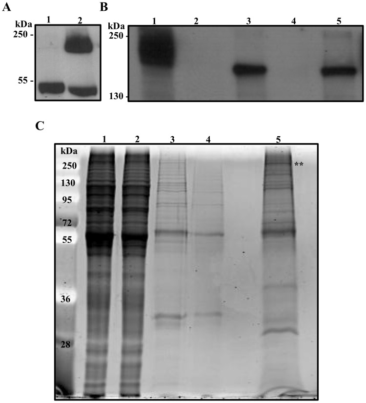
Figure 2. TAP of APC1-PTP and identification of APC1-associated proteins.
(A) Lysates of strain 427 cells transfected either with empty vector (control, lane 1) or pC.APC1.PTP (lane 2) were fractionated on 10% SDS-PAGE gel, Western blotted and simultaneously probed with anti-protein C antibody (HPC4) and anti-tubulin. The blot was then detected with an anti-mouse HRP-conjugated secondary antibody. (B) Stepwise Western blot monitoring of APC1-PTP during TAP. Samples were analyzed from 1. IgG input (1x), 2. IgG-Sepharose flow-through (1x), 3. The elute from IgG-Sepharose after TEV-protease digestion (5x), 4. The flow-through from anti-protein C matrix (5x), and 5. The EGTA final elute (20x). The blot was probed with HPC4 antibody and the values (in x) represent the relative amounts of samples analyzed. (C) Samples collected from TAP as in (B) were fractioned on 10% SDS-PAGE gel and stained with SYPRO-Ruby. (**) on the top indicates the position of APC1-PTP fusion protein.

