Figure 4.
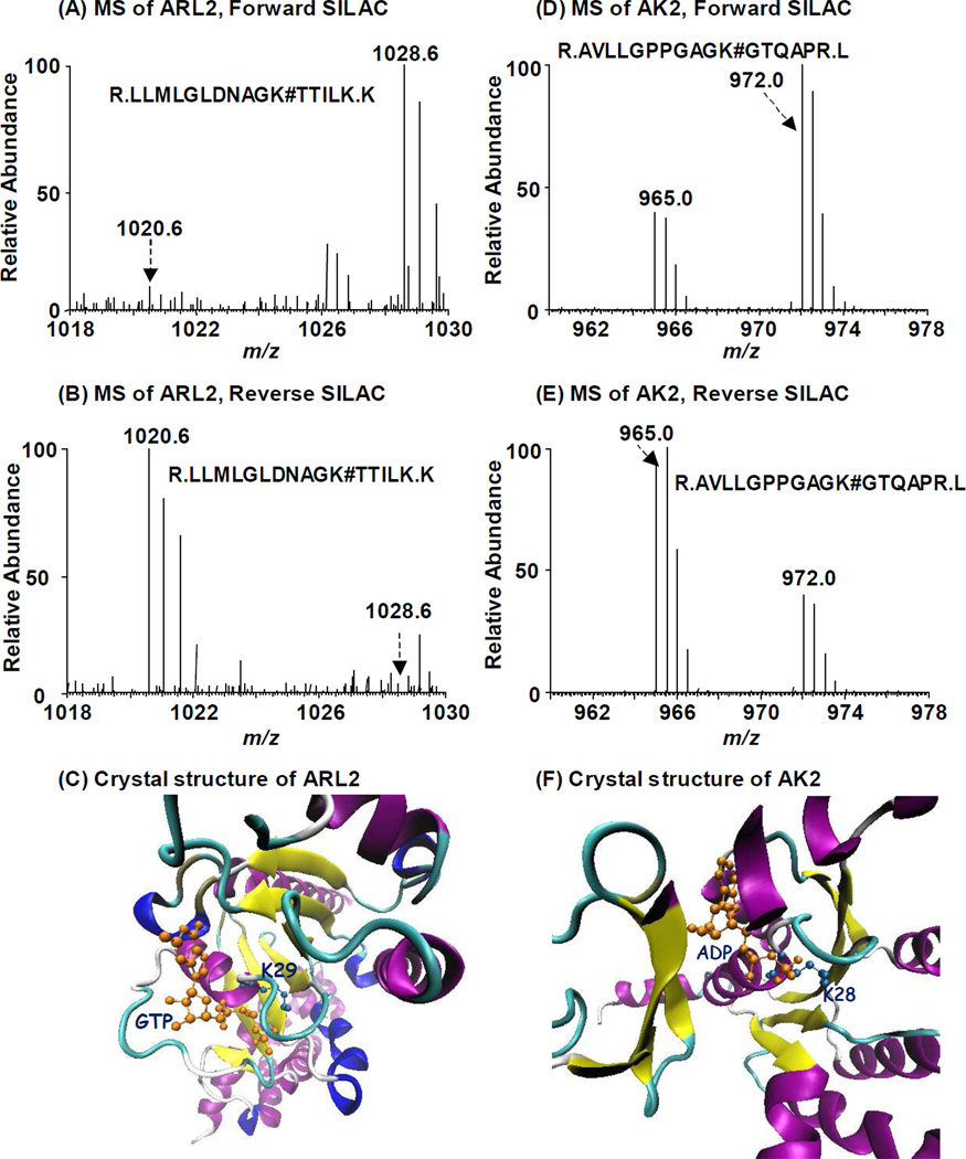
Forward- and reverse-SILAC combined with LC-MS/MS for the quantitative comparison of ATP/GTP binding affinity towards ADP-ribosylation factor-like protein 2 (A, B) and adenylate kinase 2 (C, D). # indicates the biotin-labeling site. Crystal structures of ARL2 bound with GTP (E) and AK2 bound with ATP (F) demonstrate the direct contact of nucleotide with identified lysine residues.

