Fig. 1.
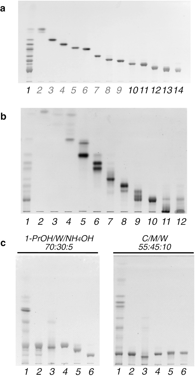
A thin-layer chromatogram shows the separation of neutral glycosphingolipids separated from the brine shrimp A. franciscana. a Fractionation by linear gradient elution. Lane 1, total neutral glycosphingolipid fraction; lanes 2–9, previously reported CMS, CDS, nAtCTS, AtCTS, nAtCTeS, AtCTeS, CPS, and CHS, respectively; lane 10, CHpS1; lane 11, CHpS2; lane 12, COS1; lane 13, mixture of COS2, CNS, and CDeS; lane 14, CDeS. b Fractionation by stepwise elution. Lane 1, total neutral glycosphingolipid fraction; lanes 2 and 3, nonpolar material fraction; lane 4, CMS; lane 5, CMS and CDS; lane 6, nAtCTS, AtCTS, and nAtCTeS; lane 7, AtCTeS; lane 8, CPS and CHS; lane 9, CHpS1, CHpS2, and COS1; lane 10, mixture of COS2, CNS, and CDeS; lanes 11 and 12, non-GSL fractions. c Purification of the fraction containing COS2, CNS, and CDeS by Iatrobeads column chromatography using ammoniacal propanol. Lane 1, total neutral glycosphingolipid fraction; lane 2, before fractionation; lane 3, non-GSL fraction; lane 4, CDeS; lane 5, CNS; lane 6, COS2. The HPTLC (a and c) and TLC (b) plates were developed in (a and b) C/M/W (60:40:10, v/v/v), (c) 1-propanol/water/ammonium hydroxide (70:30:5, v/v/v) or C/M/W (55:45:10, v/v/v). The spots were visualized by orcinol- H2SO4 reagent

