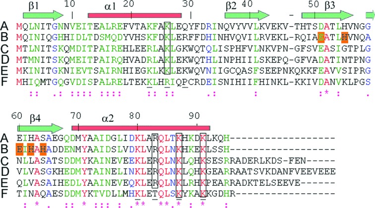
Figure 4.
ClustalW alignment. Sequences are as follows: A, E. coli HPF; B, V. cholerae HPF; C, H. influenzae YfiA; D, E. coli YfiA; E, V. cholerae YfiA; F, C. burnetii HPF. The symbols below the sequences are as follows: *, identity; :, strongly similar; ., similar; a space indicates difference. β-Strands and α-helices are indicated as transparent green arrows and red bars, respectively. The numbering scheme follows the V. cholerae HPF sequence. Underlined residues are basic residues in the C-terminal portions of α1 and α2 of YfiA of E. coli involved in the proposed ribosome-recognition site, while gray boxes highlight highly conserved residues according to Ye et al. (2002 ▶). Alignments were made using ClustalW (Thompson et al., 1994 ▶).

