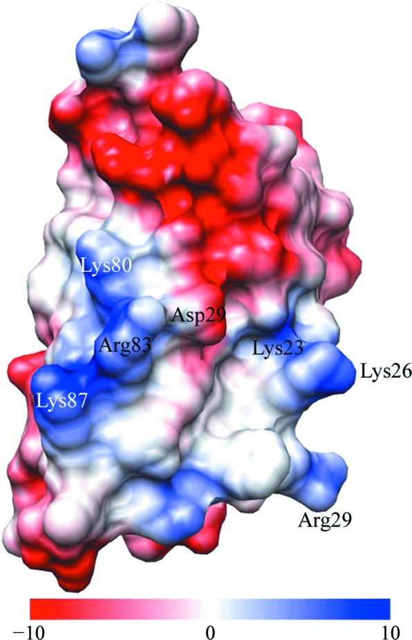
Figure 7.

Electrostatic surface map of our structure, viewing the residues of the basic patch on the surface that faces towards the ribosome. Residues in the basic patch (blue) and Asp79 (red, acidic) are indicated; the electrostatic potential is given in kcal mol−1 e−1. This figure was generated using UCSF Chimera.
