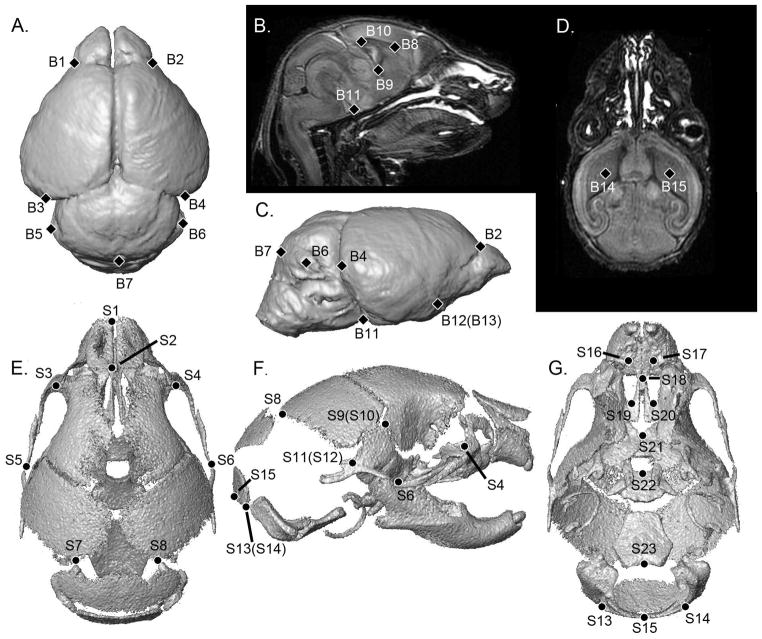
Figure 2.
Anatomical locations of the landmarks collected from the brain and skull of P0 and P2 mice, illustrated on 3-D reconstructions of MRM and μ-CT scans of a P2 unaffected mouse. A) Brain, dorsal view. B) Midsagittal slice. C) Brain, right lateral view. D) Horizontal slice. E) Skull, dorsal view. F) Skull, right lateral view. G) Skull, ventral view. Definitions for landmark abbreviations are provided in Table 1 and are further defined at www.getahead.psu.edu.

