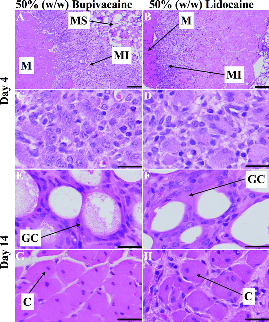
Fig. 6.
Representative light microscopy of hematoxylin/eosin-stained sections of muscles (M), and surrounding tissues at the sight of injection of 50% (w/w) bupivacaine or lidocaine loaded poly-lactic-co-glycolic acid (PLGA) microspheres (MS). (A and B) 4 days after injection, muscle injury (MI) extends deep into the muscle fascicle. (C and D) Tissue reaction was characterized by myocyte injury with abundant macrophages and occasional lymphocytes. (E and F) 2 weeks after injection, microspheres were surrounded by lymphocytes, macrophages and foreign-body giant cells (GC). (G and H) Tissue reaction was characterized by myocytes with nuclear centralization (C) surrounded by spasely distributed lymphoctes and macrophages. Scale bars 100 µm (A and B), 20 µm (C–H).

