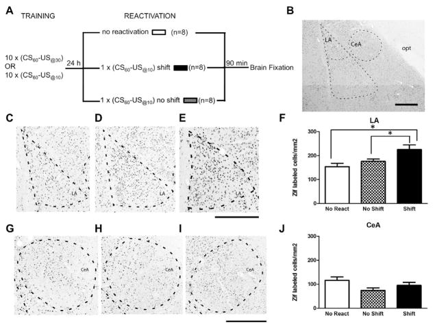
Figure 2. Memory reactivation induces synaptic plasticity in LA only when the CS-US time interval is shifted.
(A) Schematic of the experimental design. (B) Photomicrograph showing the amygdala nuclei analyzed for Zif immunoreactivity. Photomicrographs of transverse Zif-stained sections from representative cases illustrating no reactivated animals (C), no shift (D) and shift animals (E) in the LA or in the CeA (G, H, I) at 2.8mm posterior to Bregma. The broken lines delineate the nuclei borders. Abbreviations: CeA, central nucleus of amygdala, LA, lateral nucleus of amygdala; opt, optic tract. Scale bar = 200 μm. (F) (J) represent the average of Zif-648 positive cells per square millimeter (mean ± s.e.m.) across three Anterior-Posterior levels. Only the rats whose memory was reactivated with a CS-US time interval different than the one used during training showed an increase in the expression of Zif-648 positive cells in the LA (F) but not in the CeA (J). (*P<0.05 Newman-Keuls post-hoc test)

