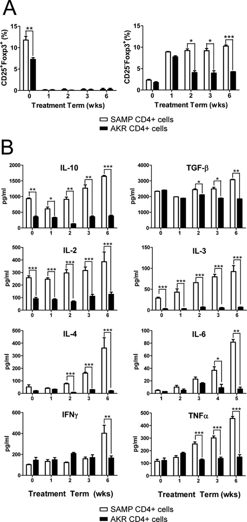
Figure 2. SAMP CD25−Foxp3+cells increase following anti-CD25 treatment and produce increased levels of effector cytokines.
CD4+ cells from anti-CD25 Ab treated SAMP or AKR mice (200µg) were isolated at 0, 1, 2, 3 and 6 weeks (n=4/time point). Mice were euthanized at 12 wks of age. (A) Time course of the proportion of CD25+Foxp3+ and CD25−Foxp3+ cells in spleen from SAMP and AKR mice. (B) Time course of Th1 and Th2 cytokines production measured by ELISA in triplicate from 72-hour cultures of SAMP or AKR unfractionated spleen CD4+ T cells (2×10⁵ cells/well) stimulated with immobilized anti-CD3 and soluble anti-CD28. Data are expressed as the mean ± SEM (*P<0.05, **P<0.01, ***P<0.001).

