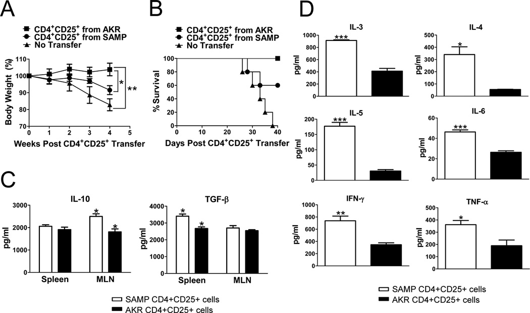
Figure 5. SAMP CD4+CD25+ cells fail to prevent the development of adoptively transfer-induced colitis.
CD4+ cells were transferred from anti-CD25 treated SAMP mice into SCID mice (6 wks, n=10) resulting in severe colitis. Six weeks later, 5 colitic SCID recipients per group received a second adoptive transfer of MLN CD4+CD25+ cells (2×10⁵) from untreated SAMP or AKR control mice (12 wks, n=3/group). The percentage of FoxP3+ cells was 90.8% and 88.3% in SAMP and AKR mice respectively. (A) Time course of body weight changes after transfer showed significant weight gain in SCID mice treated with AKR versus SAMP CD4+CD25+ cells. (B) Survival analysis of different experimental groups showed 0% survival in SAMP CD4+CD25+ treated SCID mice versus 60% in AKR CD4+CD25+ treated SCID mice. (C) Elevated levels of secreted IL-10 and TGF-β measured from 3-day cultures of SAMP (n=4 mice) or AKR (n=6 mice) MLN or spleen CD4+CD25+ T cells (2×10⁵ cells/well) stimulated with immobilized anti-CD3 and soluble anti-CD28. (D) TNF-α, IFN-γ, IL-3, IL-4, IL-5, and IL-6 measured from 3-day cultures were elevated in SAMP (n=4 mice) compared to AKR (n=6 mice) MLN or spleen CD4+CD25+ T cells (2×10⁵ cells/well) stimulated with immobilized anti-CD3 and soluble anti-CD28. Data are expressed as the mean ± SEM (*P<0.05, **P<0.01, ***P<0.001).

