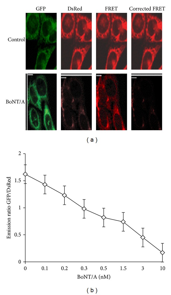
Figure 3.

(a) Measuring toxin sensor in living cells. The stable clone 5A3 expressing the AcGFP-SNAP-25-DsRED sensor was treated with 10 nM BoNT/A holotoxin, and the FRET signals of 100 cells were analyzed after 72 hours. (as described in Materials and Methods). Control cells not treated with toxins were analyzed in parallel (upper panel). Images of representative cells are shown. This sensor yielded significant FRET (upper “corrected FRET”), which was abolished after cells were treated with BoNT/A (72 h, lower “corrected FRET”). Three images (GFP, FRET, and DsRED) were taken for each set of cells sequentially, using exactly the same settings. FRET signal was measured by exciting AcGFP and detecting DsRED signal. (b) Measurements of C-terminal reporter fragment degradation in clone 5A3 using a microplate reader (BioTek). The GFP and DsRED emissions were collected by fluorescence microplate reader (BioTek). Emissions were plotted as a function of BoNT/A at 0.03, 0.05, 0.15, 0.3, 1.5, 3.0, and 10 nM concentrations. Data are presented as means ± SD (n = 3).
