Figure 4.

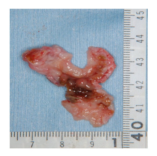
The excised tumor. It was milky-white in colour, with a slightly rough surface. The transverse section was largely cystoid, but solid portions were also observed.

The excised tumor. It was milky-white in colour, with a slightly rough surface. The transverse section was largely cystoid, but solid portions were also observed.