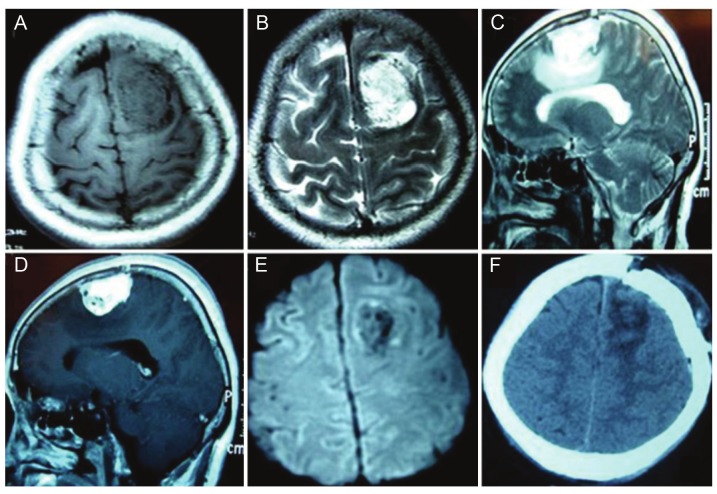
Figure 2.
Neuroimaging for an angiomatous meningioma at left frontal region a. A. Axial T1WI: Hypointense masswith the voids of blood vessels. B. Axial T2WI: Hyperintense mass with the voids of blood vessels. C. Sagittal T2WI:Hyperintense mass with peritumoral edema. D. Sagittal postcontrast T1WI: Tumor with homogeneously enhancementwith the voids of blood vessels. E. DWI: Hypointense and no restriction with the voids of blood vessels. F.Follow-up CT scan: Gross total resection without evidence of recurrence.

