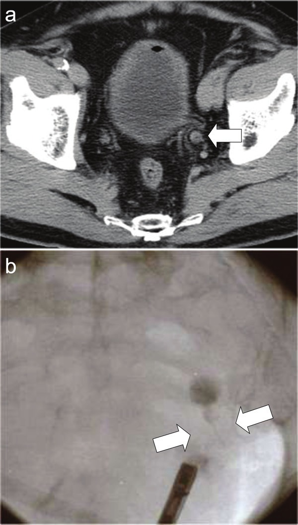
Figure 1.

Radiological images of the ureteral tumor. A: Computed tomographic image of the pelvis demonstrating the tumor in the lower portion of the left ureter (arrow). B: Retrograde urographic image revealing an obstruction of the lower portion of the left ureter (arrows).
