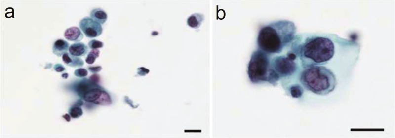
Figure 2.

Photomicrographs of cells from the voided urine. A: Degenerating, incohesive atypical cells of various sizes containing hyperchromatic nuclei of various sizes are present (Papanicolaou stain; the scale bar indicates 10 μm). B: Neoplastic cells exhibit a relatively rich cytoplasm and a marginally situated nucleus containing fine or coarse granular hyperchromatic material and an enlarged nucleolus (Papanicolaou stain; the scale bar indicates 10 μm).
