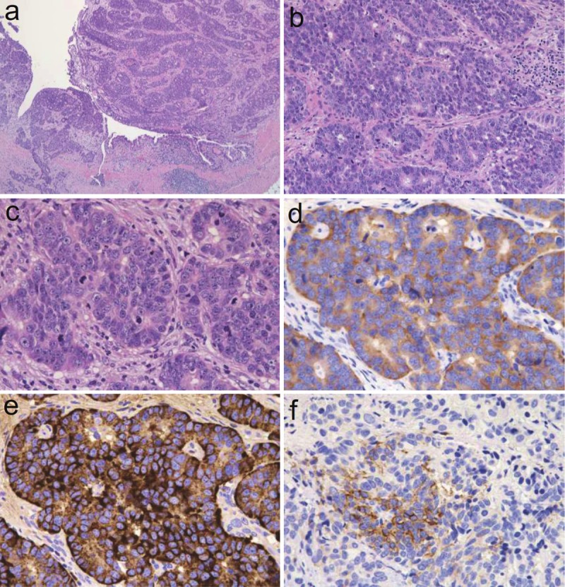
Figure 4.

Photomicrographs of the ureteral tumor. A: Low-power view of neoplastic cells in irregularly shaped nests and neoplastic cells that penetrated the muscular layer (hematoxylin and eosin stain, objective 20x). B: Medium-power view of neoplastic cells arranged in rosette-like, palisading, or organoid patterns (hematoxylin and eosin stain, objective 20x). C: High-power view of neoplastic cells harboring a rich cytoplasm with large nuclei containing fine or coarse granular hyperchromatic material and nuclei and moderately enlarged nucleoli. These cells are accompanied by large numbers of mitotic cells and apoptotic bodies (hematoxylin and eosin stain, objective 20x). D: Neoplastic cells positive for the expression of synaptophysin (immunohistochemistry, objective 40x). E: Neoplastic cells positive for the expression of chromogranin A (immunohistochemistry, objective 40x). F: Neoplastic cells positive for the expression of CD56 (immunohistochemistry, objective 40x).
