
Figure 5.
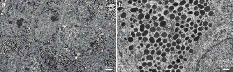
Transmission electron micrographs of the ureteral tumor. A: Neoplastic cells harboring abundant dense granules in a relatively rich cytoplasm (the scale bar indicates 2 μm). B: The cytoplasm of a neoplastic cell containing numerous dense granules ranging in size from 200 to 400 nm in diameter (the scale bar indicates 500 nm).