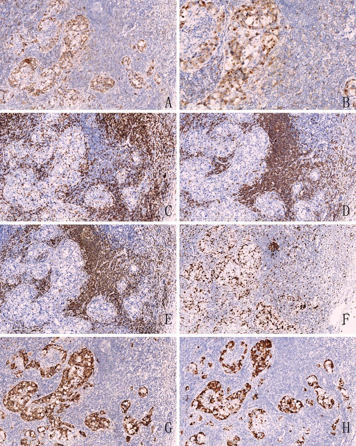
Figure 2.
Immunohistochemical analysis. A. The tumor cells were positive for cytokeratin(pan), roblastic reticulum cells showed a typical dendritic shape (Original magnification ×100). B. High magnification showed that positive reactivity of cytokeratin(pan) with Golgi-associated dot-like staining pattern. (Original magnification ×200). C. The tumor cells were negative for CD3, but T lymphocytes showed a nuclear staining (Original magnification ×100). D. CD20, E. Pax-5 were also negative in atypia tumor cells and positive for B lymphocytes. (Original magnification ×100). F. showed the high Ki-67 labeling index. (Original magnification ×100). G. The large lymphocytes are strongly CD30 positive with a membrane and Golgi pattern of staining. (Original magnification ×100). H. ALK/P80 immunohistochemistry shows strong cytoplasmic staining in the neoplastic cells. (Original magnification ×100).

