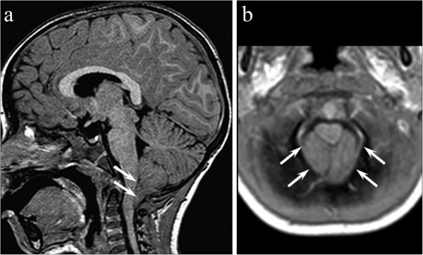
Figure 2.

Sagittal contrast-enhanced T1-weighted (a) showing a caudal protrusion (12 mm) of the elongated triangular-shaped tonsils below the foramen magnum (white arrows). Axial T1-weighted image (b) confirms cerebellar tonsils filling the foramen magnum and effacing the cysterna magna (white arrows).
