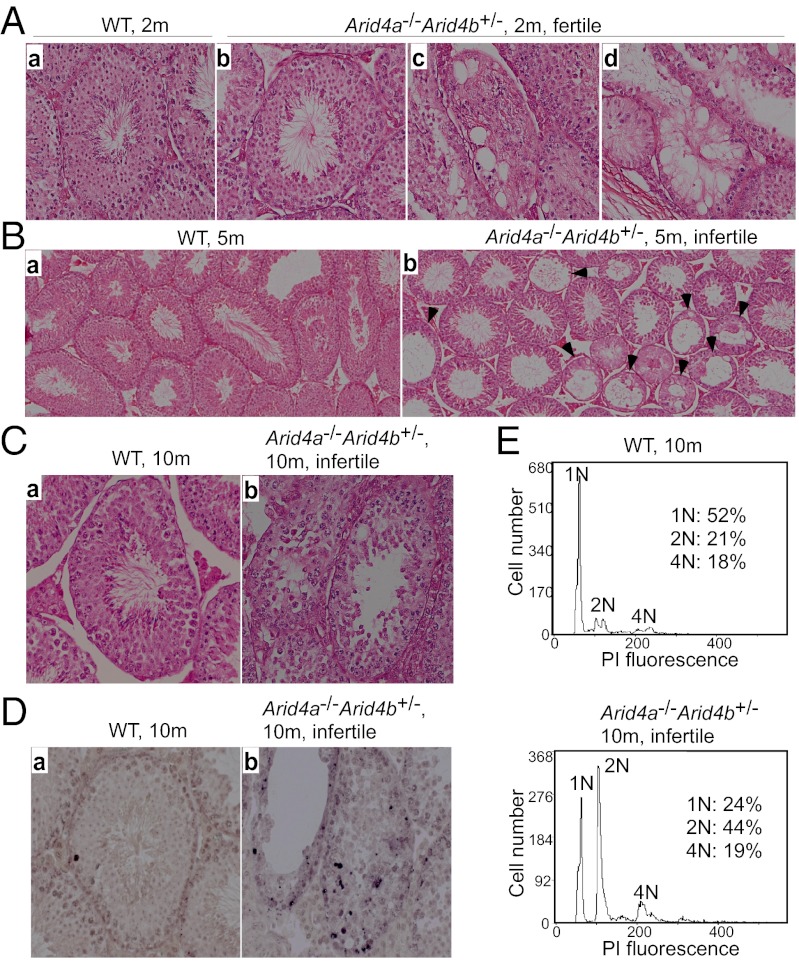
Fig. 3.
Deterioration of seminiferous tubules and arrest of spermatogenesis in the Arid4a−/−Arid4b+/− testes. (A) H&E-stained sections of seminiferous tubules of testes from wild-type (a) and fertile Arid4a−/−Arid4b+/− (b–d) mice at 2 mo of age. Original magnification was 400× . (B and C) H&E-stained sections of wild-type (a) and infertile Arid4a−/−Arid4b+/− (b) testis from mice at 5 mo of age (B) and at 10 mo of age (C). Original magnifications were 200× (B) and 400× (C). Black arrowheads indicate loss of architecture of the seminiferous tubules in the Arid4a−/−Arid4b+/− testes (B, b). (D) In situ TUNEL assay of testis sections from wild-type (a) and infertile Arid4a−/−Arid4b+/− (b) male mice at 10 mo of age. Apoptotic cells were stained brown. Original magnification was 400× . (E) Flow cytometric analysis of propidium iodide fluorescence-stained DNA of testicular cells from wild-type (Upper) and infertile Arid4a−/−Arid4b+/− (Lower) male mice at 10 mo of age.

