Fig. 6.
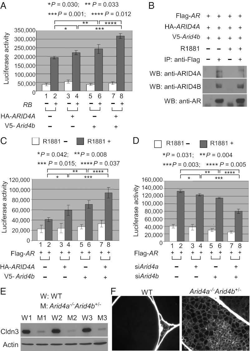
Arid4a and Arid4b regulate Cldn3. (A) ARID4A and Arid4b induce RB activity on the Cldn3 promoter. TM4 cells were transfected with the Cldn3 promoter-regulated luciferase reporter along with the expression vectors for RB, HA-ARID4A, or V5-Arid4b as indicated. Luciferase activity was measured 48 h after transfection. Shown are means ± SD from three experiments performed in triplicate. (B) ARID4A and ARID4B were associated with AR in a ligand-independent manner. TM4 cells were transfected with the expression vectors for Flag-AR, HA-ARID4A, and V5-Arid4b. After the transfected cells were treated with or without R1881 (100 nM), immunoprecipitation experiments were performed using the anti-Flag Ab, and then Western blot analyses were performed by using antibodies against ARID4A, ARID4B, or AR. (C) ARID4A and ARID4B additively induce AR activity on the Cldn3 promoter. TM4 cells were transfected with the Cldn3 promoter-regulated luciferase reporter along with the expression vectors for Flag-AR, HA-ARID4A, or V5-Arid4b as indicated. The transfected cells were treated with or without R1881, and luciferase activity was measured thereafter. Shown are means ± SD from three experiments performed in triplicate. (D) Knockdown of Arid4a and Arid4b synergistically reduced the AR activity on the Cldn3 promoter. TM4 cells were first transfected with siArid4a and/or siArid4b. The next day, these cells were transfected with the Cldn3 promoter-regulated luciferase reporter and the expression vector for AR. After the cells were treated with or without R1881, luciferase activity was determined. Shown are means ± SD from three experiments performed in triplicate. (E) Decreased expression of Cldn3 in the testes of the Arid4a−/−Arid4b+/− mice was validated at the protein level by Western blot analysis. Actin was used as a loading control. Cldn3, 23 kDa; actin, 47 kDa. (F) Increased permeability of the blood–testis barrier in the Arid4a−/−Arid4b+/− testes. The permeability of the blood–testis barrier was assessed by injection of a biotin tracer into testes of the Arid4a−/−Arid4b+/− and wild-type mice at 3 mo of age. The biotin tracer was detected by Alexa Fluor 488 streptavidin. Biotin is present in the adluminal space of the Arid4a−/−Arid4b+/− testes.

