Fig. 3.
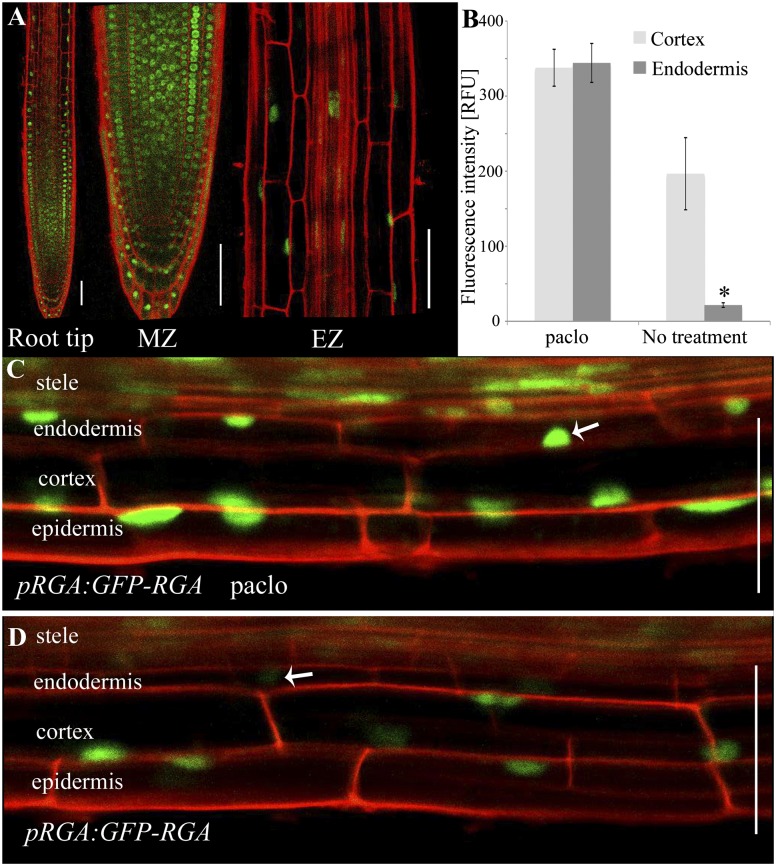
GFP-RGA signal is reduced in elongating endodermal cells. (A) GFP-RGA localization in the root. MZ, meristematic zone; EZ, elongation zone. (B) Relative fluorescence intensity of GFP-RGA in the cortex and endodermal nuclei of the elongation zone with and without 2 M Paclo treatment. Shown are averages ± SE (three root images, three cells/image, three sampling points/cell; n = 27). (C and D) Images of the elongation zone showing GFP-RGA levels (C) with (D) without Paclo treatment. White arrows indicate the endodermal cell nuclei. *Significantly different relative to the respective cortex cell at P ≤ 0.001 by Student t test. (Scale bars, 50 μm.)

