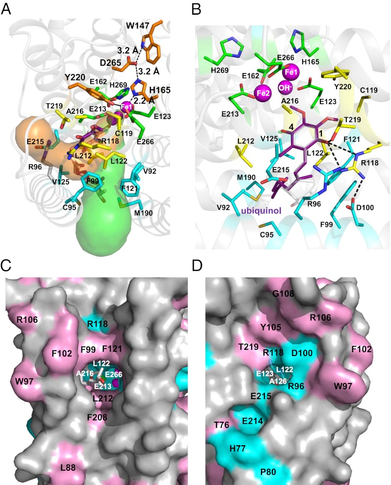
Fig. 5.
Putative ubiquinol binding cavities in TAO. (A) Two hydrophobic cavities predicted by CAVER protein-analysis software (38). The bound AF2779OH (isoprenoid tail) occupies the green cavity. Putative residues involved in electron transfer are shown as orange sticks. (B) Ubiquinol binding model predicted by the superposition of a ubiquinol molecule (purple stick) onto the bound AF2779OH (translucent white stick) of the TAO–AF2779OH complex. Magenta spheres are diiron (Fe–OH−–Fe), the green stick represents residues coordinating to diiron, and yellow and cyan sticks are residues interacting with the aromatic head and isoprenoid tail, respectively. Hydrogen bonds are depicted as dotted lines. Surface views of the (C) green and (D) orange cavities shown in A. Cyan and pink colors stand for conservation of AOX residues in all eight and over four organisms in SI Appendix, Fig S5, respectively.

