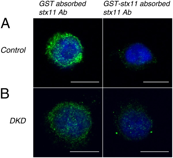
Fig. 2.
Confocal immunofluorescence microscopy revealing that the subcellular localization of syntaxin-11 is unaltered on Munc18-1/2 DKD. Control and stable Munc18-1/2 DKD RBL-2H3 cells were permeabilized and stained with either GST or GST-syntaxin-11–absorbed rabbit polyclonal anti-syntaxin-11 antibody followed by Alexa488-conjugated goat anti-rabbit antibody (Invitrogen) and DAPI (see SI Materials and Methods for more detail). Green indicates syntaxin-11 and blue indicates DAPI. (A) Control and (B) Munc18-1/2 DKD cells. Note that there is substantially diminished green intensity (Right) where GST-syntaxin-11 absorbed the anti-syntaxin-11 antibody was used to stain. (Scale bar, 10 μm.)

