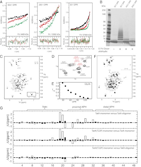
Fig. 1.
(A) Analytical ultracentrifugation of 167 µM TatA at DPR of 604:1, 250:1, and 30:1. Molecular weights of 6,489 kDa, 13,582 kDa, and 57,484 kDa, respectively, were determined from simultaneous fitting to data at rotor speeds of 20 K (black), 30 K (red), and 40 K (green). The monomeric TatA molecular mass was calculated to be 6.2 kDa under analytical ultracentrifugation conditions (50% 2H2O), indicating average oligomeric states of 1.0, 2.2, and 9.3, at DPR of 604:1, 250:1, and 30:1, respectively. (B) SDS/PAGE of TatA (500 µM) cross-linked with glutaraldehyde at DPR of 30:1, 425:1, and 850:1. (C) 1H-15N HSQC of monomeric TatA (500 µM) at a DPR of 250, with resonance assignments indicated. Resonances arising from the C-terminal 6-His tag are indicated with an asterisk. (D) 1H-15N HSQCs of the glycine region of TatA at an intermediate DPR of 60. TatA at DPR of 60 shows peak doubling for Gly26, Gly29, and Gly33 corresponding to monomeric and oligomeric TatA molecules. (E) The oligomeric fraction of the total peak volume from the two resolved Gly26 peaks: folig = [volumeoligomer/(volumeoligomer + volumemonomer)] as a function of DPR. (F) 1H-15N SOFAST-HMQC of oligomeric TatA (500 µM) (DPR = 30) with resonance assignments indicated. (G) Chemical shift differences in 1H (open bars) and 15N (filled bars) between the indicated TatA constructs.

