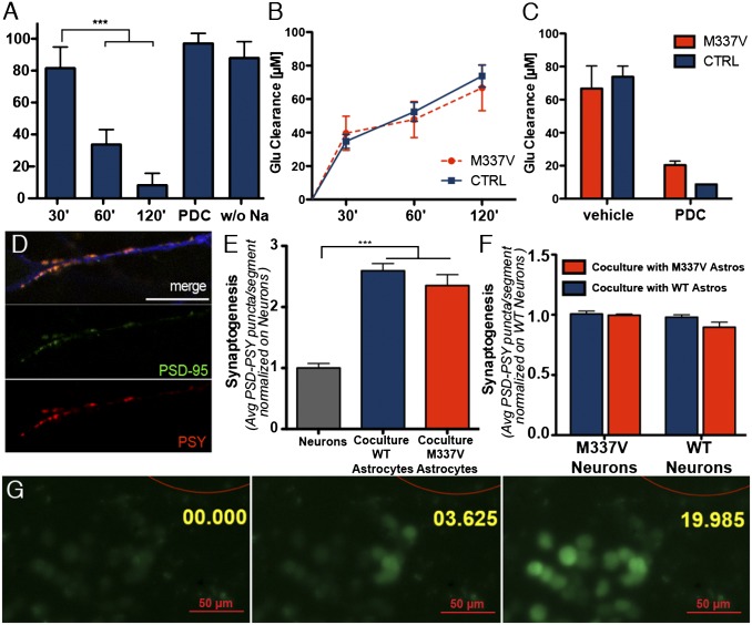
Fig. 2.
Functional characterization of iPSC-derived astrocytes. (A) l-glutamate uptake assay on iPSC-derived astrocytes. iPSC-derived astrocytes cleared l-glutamate in a time-dependent fashion. ***P < 0.01 (one-way ANOVA). The uptake was abolished by either the absence of sodium or the presence of 2 mM L-trans-pyrrolidine-2,4-dicarboxylic acid (PDC; both negative CTRLs were run for 120 min). (B) M337V iPSC-derived astrocytes did not show a significant difference in glutamate clearance rate, compared with CTRL astrocytes (one-way ANOVA; P > 0.05 for all times considered). (C) Glutamate uptake fold increase. M337V and CTRL astrocytes were able to clear >75% of l-glutamate from the medium in 120 min, and uptake was inhibited equally by PDC. No significant difference was observed between the groups. (D) Representative immunofluorescence panels from synaptogenesis assay. Image shows representative neurite segment used for analysis, stained with TuJ1 (blue), PSY (red), and PSD-95 (green; scale bar: 15 µm). (E) iPSC-derived astrocytes significantly promoted synaptogenesis when cocultured with differentiating neurons. ***P < 0.01 (one-way ANOVA). The increase in synaptogenesis was measured by scoring the number of PSY+/PSD-95+ puncta in 30-µm neurite segments (coculture M337V, 2.4 ± 0.2, SEM; coculture WT, 2.6 ± 0.1, SEM; neurons, 1.00 ± 0.08, SEM). No significant difference was observed in the behavior of M337V or CTRL iPSC-derived astrocytes. Data represent the average of three independent experiments, each with two clones of M337V (M337V-1 and -2) iPSC and two independent CTRL lines (CTRL-1 and -2); data from different lines were pooled for graphical presentation. (F) Synaptogenesis assay with M337V or WT patterned neurons revealed that astrocyte promotion of synapse formation was independent from the genotype of cocultured neurons and that M337V astrocytes do not exert a negative effect on synaptic formation, even to mutant neurons. (M337V neurons on WT astrocytes, 0.90 ± 0.04, SEM; M337V neurons on M337V astrocytes, 0.98 ± 0.03 SEM; WT neurons on WT astrocytes, 1.00 ± 0.01 SEM; WT neurons on M337V astrocytes, 1.01 ± 0.02, SEM). (G) iPSC-derived astrocytes propagate calcium waves to adjacent cells upon mechanical stimulation. Red outline represents glass bead used for stimulation (200-µm diameter). Annotation represents time in seconds from stimulation.

