
Fig. 4.
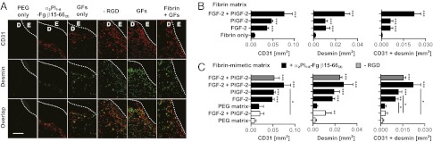
Angiogenesis within the granulation tissue. Angiogenesis was revealed by staining for endothelial (CD31+) cells and smooth muscle (desmin-positive) cells. (A) Representative images are shown. GFs, FGF-2 + PlGF-2. E, epidermis; D, dermis. The hashed line indicates the basement membrane. (Scale bar: 0.2 mm.) (B and C) The graphs show quantification of stained areas for CD31 and desmin as well as the overlay (n ≥ 4; mean ± SEM). *P < 0.05; **P < 0.01; ***P < 0.001, Student t test.